
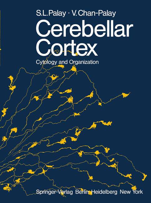
Cerebellar Cortex
☆☆☆☆☆
(0 opinii)
Najniższa cena (z dostawą)
20 034,00 JPY
Typowa cena3 050,57 PLN
Najniższa (90 dni)145,59 PLN
Liczba ofert6
Ostatnia aktualizacja1 tydzień temu
Historia ceny (90 dni)
Pełna historia
2026-08-08
2026-08-15
| Zaktualizowano | Cena |
|---|---|
| 2026-08-08 | 145,59 |
| 2026-08-15 | 145,59 |
| Sprzedawca | Cena produktu | Dostawa | Razem | Dostępność | Aktualizacja | |
|---|---|---|---|---|---|---|
| SP SpringerNatureLink Shop INT | 20 019,00 JPY | 15,00 JPY | 20 034,00 JPY | Dostępny | 5 dni temu | Zobacz ofertę |
| SP Springer Nature Author | 20 019,00 JPY | 25,00 JPY | 20 044,00 JPY | Dostępny | 1 tydzień temu | Zobacz ofertę |
| SP SpringerNatureLink Shop INT | 139,99 USD | 19,00 USD | 158,99 USD | Dostępny | 5 dni temu | Zobacz ofertę |
| SP SpringerNatureLink Shop INT | 159,99 USD | 0 zł | 159,99 USD | Dostępny | 5 dni temu | Zobacz ofertę |
| SP SpringerNatureLink Shop INT | 159,99 USD | 15,00 USD | 174,99 USD | Dostępny | 5 dni temu | Zobacz ofertę |
| SP SpringerNatureLink Shop INT | 165,50 EUR | 25,00 EUR | 190,50 EUR | Dostępny | 5 dni temu | Zobacz ofertę |
Ceny i dostępność mogą ulec zmianie. Ostatnia aktualizacja: 08.08.2026 23:32.
0,0
☆☆☆☆☆
0 opinii
5★
0%
4★
0%
3★
0%
2★
0%
1★
0%
Opinie o produkcie
Brak opinii — bądź pierwszy!
The origins of this book go back to the first electron microscopic studies of the central nervous system. The cerebellar cortex was from the first an object of close study in the electron microscope, repeating in modern cytology and neuroanatomy the role it had in the hands of RAMON y CAJAL at the end of the nineteenth century. The senior author vividly remembers a day early in 1953 when GEORGE PALADE, with whom he was then working, showed him an electron micrograph of a cerebellar glomerulus, saying "That is what the synapse should look like. " It is true that the tissue was swollen and the mitochondria were exploded, but all of the essentials of synaptic structure were visible. At that time small fragments of tissue, fixed by immersion in osmium tetroxide and embedded in methacrylate, were laboriously sectioned with glass knives without any predetermined orientation and then examined in the electron microscope. After much searching, favorably preserved areas' were studied at the cytological level in order to recognize the parts of neurons and characterize them. Such procedures, dependent upon random sections and uncontrollable selection by a highly erratic technique of preservation, precluded any systematic investigation of the organization of a particular nucleus or region of the central nervous system. It was difficult enough to distinguish neurons from the neuroglia.
Podobne produkty
 50 Years of Philosophy and Medicine
od $107.30
50 Years of Philosophy and Medicine
od $107.30
 Understanding Cybersecurity Law and Digital Privacy
od $122.63
Understanding Cybersecurity Law and Digital Privacy
od $122.63
 Decisions in International Trade and Logistics
od $122.63
Decisions in International Trade and Logistics
od $122.63
 Advances in Economic Measurement
od $122.63
Advances in Economic Measurement
od $122.63
 Nanomaterials
od $137.96
Nanomaterials
od $137.96
 Sustainable Scaffolds-based Strategies in Tissue Engineering and Regenerative Medicine
od $162.87
Sustainable Scaffolds-based Strategies in Tissue Engineering and Regenerative Medicine
od $162.87
 Narratives in Educational Research
od $162.87
Narratives in Educational Research
od $162.87
 Environmental Biology of the Young Chinese Sturgeon
od $210.78
Environmental Biology of the Young Chinese Sturgeon
od $210.78